Image contest 2017 – The winners!
Light microscopy
[tabs]
[tab title=”The winner”]
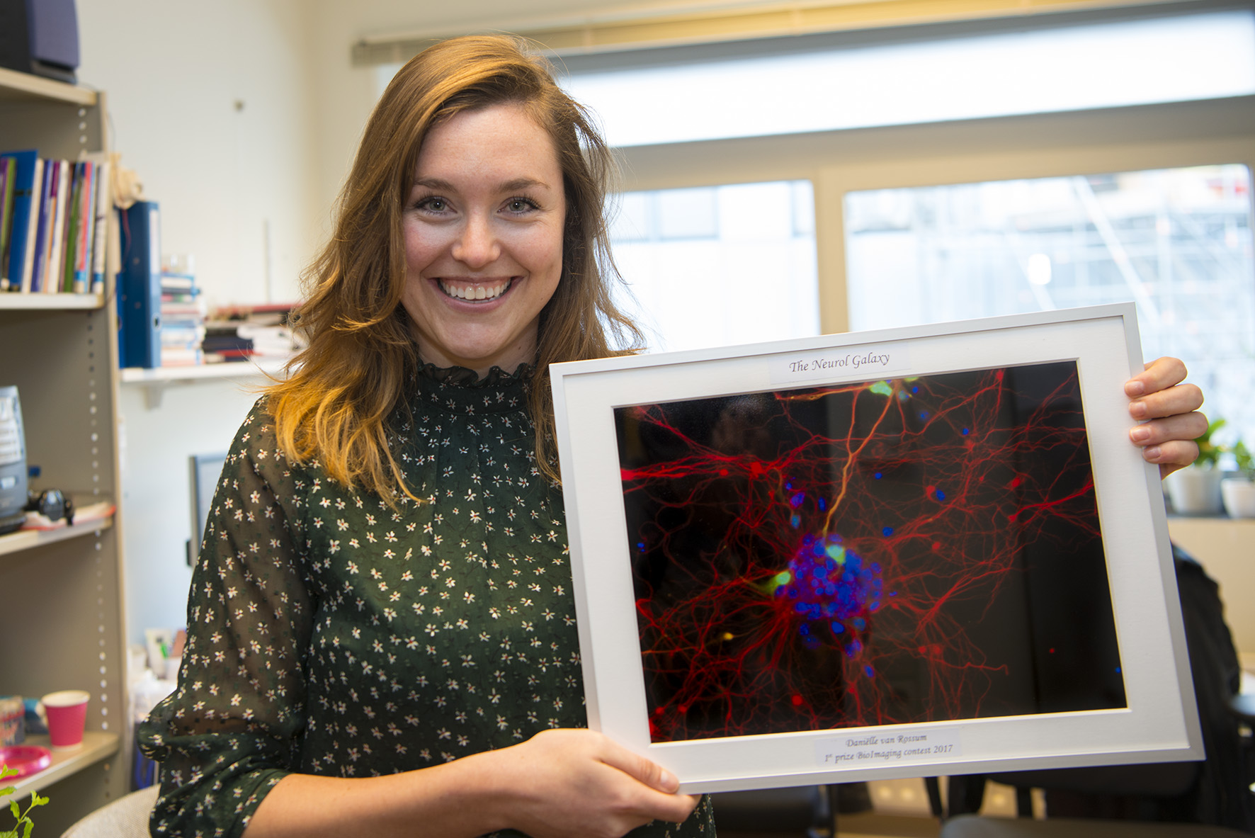
 Danielle van Rossum, UMC
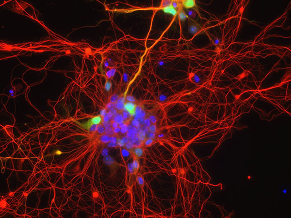
The artistic name of the image: The neuronal galaxy
Description: The picture originates funnily enough from a failed experiment where I tried to culture single cell cortical mouse neurons in vitro. The single cell part didn’t turn out so well and resulted in a clump of neurons together forming a complex network. This ‘network’ combined with fluorescent imaging resulted in what I call a “neural galaxy” phenotype as it reminds me of the complexity, beauty and mystery of the galaxy with a touch of Star Wars lightsaber effect.
Microscope: Zeiss LSM 880 confocal
Printed!:
[/tab]
[tab title=”2nd prize”]
 Simone Kersten, UMC
Artistic title: Luminosity
Description: A two-dimensional representation of a three-dimensional small intestinal mouse crypt, based on a simple temporary depth colour coding of a nuclear staining (Dapi). This ultimately results in a magnificent and colorful image that evokes our curiosity and imagination.
Microscope: Leica TSC SP8 X confocal microscope
[/tab]
[tab title=”3rd prize”]
 Lars-Eric Fielmich, UU
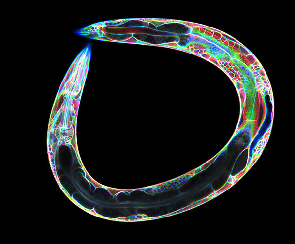
Artistic title: Psychadelegans
Scientific description: PH-eGFP marks all the cell-membranes in a Caenorhabditis elegans larva.
Microscope: Spinning Disc microscope
[/tab]
[/tabs]
Electron microscopy
[tabs]
[tab title=”The winner”]
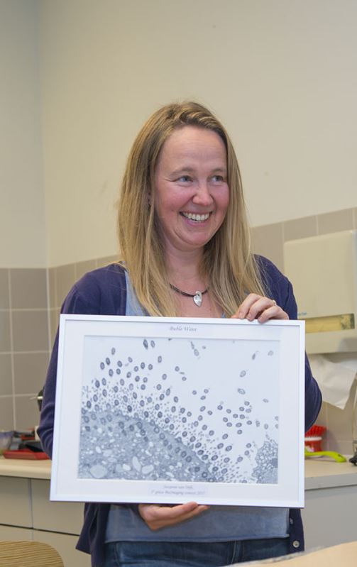
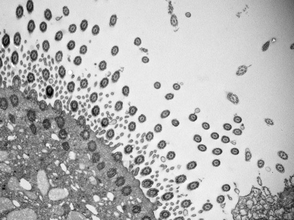 Suzanne van Dijk, UMC
Artistic title: Bubble Wave
Microscope: FEI Tecnai 12
Scientific description: Cross sections of cilia in mouse trachea.
Printed!:
[/tab]
[tab title=”2nd prize”]
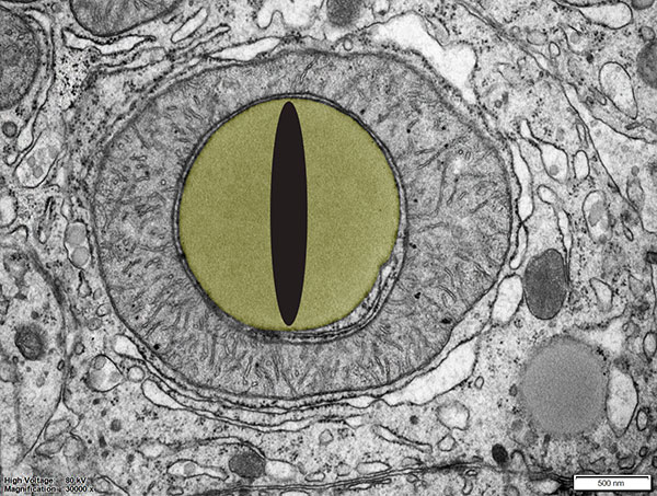 Cilia de Heus, UMC
Artistic title: Evil mitochondria eye
Microscope: Jeol1010
Scientific description: Mice liver with WASH_KO; It is a mitochondria with a lipid droplet
[/tab]
[tab title=”3rd prize”]
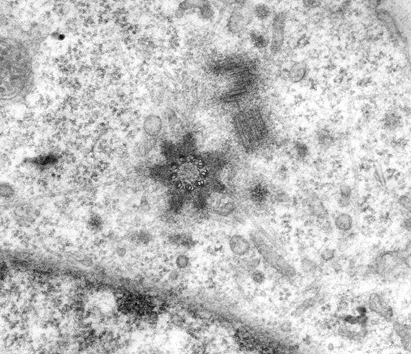 Ann De Mazière, UMC
Artistic title: Good Morning Starshine!
Microscope: Jeol 1010
Scientific description: It shows a cross-section of the basal body of a primary cilium in the centrosome of a mammalian cell. Radiating from this basal body, the basal feet with attached microtubules are visible.
[/tab]
[/tabs]